
Soravis Prakkamakul on Facial Perception
Soravis Prakkamakul, a graduate student at the School of Information, received a Spring 2018 BCNM Conference Grant to help cover his costs attending the 63rd Plastic Surgery Research Council (PSRC) annual meeting in Birmingham, Albama. Read more about his experience in his own words below!
On Saturday, May 19th, 2018 I’ve got an opportunity to present my paper “Reflexive Visual Inspection of Cleft Lip Faces – Analysis of Lookzone Focus Over Time” at the 63rd Plastic Surgery Research Council (PSRC) annual meeting in Birmingham, Albama. First, I would like to thank the Berkeley Center for New Media for giving me the opportunity. As the name suggested, it is a medical conference, and I was very excited to be one of the few information science practitioners there. Attending the meeting was an experience that was very different from my work at I School and it is always interesting to learn things happening in other fields. Most importantly, the experience suggests some ideas on the gaps in the industry that could be fulfilled by information technology and human-centered design.
Overview of the research
Human reflexively inspects faces, and facial appearance can affect the quality of life in so many ways. Our research team, consisting of Dr. Thanapoom Boonipat from Mayo Clinic, Professor Kevin Flemming from Norwich University, VT, and our PI, Dr. Mitchell Stotland from Sidra Medicine, Qatar, was interested in how people perceive the faces with facial deformity by using eye tracking data. Before I came on board, the group’s past research explored the distribution of time spent on each facial regions, the effect of surgical reconstruction on observation, and the effect of observer’s social and cultural background.
However, what has never been reported before is the aggregated observation patterns over time (e.g. where do people focus first, and for how long). We conducted an eye tracking study where 402 subjects observed a slide deck containing images randomly assigned from a group of 179 experimental and 179 control facial obtained from the senior author's practice. Experimental images included 41 individuals with repaired cleft lip. Twenty standardized lookzone regions were mapped onto each facial image. My role was to extract interesting insights on the temporal observation pattern of cleft faces using state sequence analysis and data visualization. Some examples of our visualizations can be seen in Figure 1.
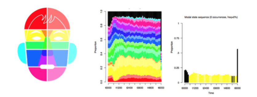
Figure 1: Color coding system (left), look zone distribution over time (center),
the most representative look zone over time (right)
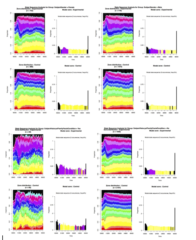
Our results showed that observers focused on the deformed regions early on in their observation then revert back to the normal pattern around the periorbital region. Males maintain focus on cleft defects twice as long than females. Personal and family history of facial deformity result in dramatic increase in focus on cleft deformed regions of the face. And, bilateral clefts were less of a visual draw than unilateral clefts.
The results could add to the understanding on the early visual processing of facial deformity. This information could be used as an objective metrics on the outcome of surgical reconstruction and could also help prioritize different surgical tasks.
Interesting Works
Although there are many exceptional medical research presentations going on at the meeting, my attention went towards papers that made innovative use of information and fabrication technology. One interesting area is the use of fabricated materials for medical scaffolding. A group of researchers explored the use of 3D-printed bioactive ceramic for bone tissue scaffolding. Usually, artificial materials could inhibit the growth of tissues, but the group proposed an approach of coating the scaffold in osteogenic agent dipyridamole along with presenting the optimal concentration of the coat. Another group presented a scaffolding method for ear reconstruction which is very challenging as engineered ear tissues usually lost shape as they grow. The group proposed a fabrication method of the external ‘cage’ that would control the contour of the growing tissue.
Another group of works went towards novel methods for the evaluation of surgery results. A group of researchers from UC Irvine Department of Plastic Surgery built a convolutional neural network that outperformed residents at classifying breast augmentation. There are also works at the intersection of plastic surgery and human-centered design. A group of researchers from University of Michigan and Ohio State University designed and tested a web-based risk calculator to improve patient decision making for breast reconstruction.
Overall, advancements in plastic surgery research involve envisioning the shape and form of reconstructions, controlling the physical shape of organic structures, and effectively evaluating the surgical outcomes. For me, fabrication technology, data science, and human-centered design hold huge potential for such areas and it is exciting to see the examples of such intersection coming alive at this conference.